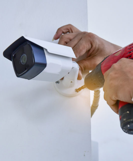
What Is Angiography in the Field of Medicine?
Angiography is quite a well-known machine. It makes us think of a complex graph machine, that shows lines going up and down to follow the rhythm of the heart. Most of us don’t know how to read these types of graphs, although we do know that a straight line means that someone has died. It may surprise you, however, that this is not at all what angiography is. Rather, angiography is a collective term for all types of medical photography, including the various machines offered through MedPhotoManager.com. Medically speaking, it is a form of imaging technique, therefore. Specifically, all tools classified as angiography tools use x-ray technology to examine the heart chambers and the blood vessels, including the arteries and veins.
Understanding Angiography
Angiography stems from the Ancient Greek word “angeion”, which translates as “vessel”, and the word “graphein”, which translates as “to record or write”. The image that is produced through an angiography is known as an angiogram or angiograph. The process of producing this image is known as arteriography or angiography. It is a technique that was developed by Antonio Egas Moniz, a Portuguese neurologist, in 1927.
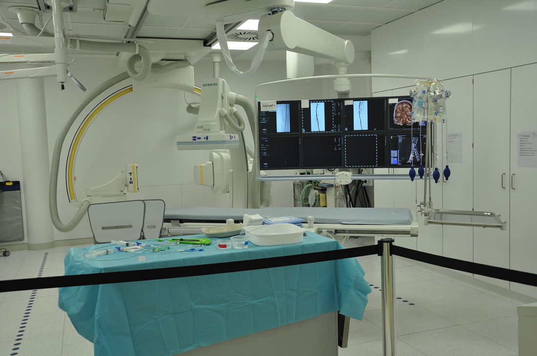
A coronary angiography is a process. First, a catheter tube or wire is inserted into the main artery of the thigh or arm. It is then placed near the opening of the arteries that provide blood supply to the heart, or near the heart itself. A contrast medium, or dye, is then injected, which can be seen through x-ray technology. This creates the picture that forms the angiogram. If the coronary tubes fill completely with the dye, then there is no blockage in the arteries.
The catheter’s tip is able to reach the coronary tube through a process of trial and error, which makes this a very delicate procedure. The fluoroscopy monitor, with heavy radiation, shows where the tip is, enabling the physician to move it towards the right location. This is dangerous, however, as a small error can lead to the tubes and arteries being punctured. Of course, there are always risks with any medical procedure, including the risk of pain and infections. Hence, if this procedure is offered to someone, it is because a physician believes the benefits outweigh the risks.
Complication rates during an angiography are around 1 for every 1,000 patients. In less than 1% of patients, major vascular complications, serious ventricular arrhythmia, stroke, myocardial infarction, or death occurs. More common complications include pericardial effusions, hypotension, blood clots, kidney damage, and cardiac arrhythmias. While more common, they are still rare. Furthermore, modern medical science can resolve these problems if need be.
The angiography has been around for nearly 100 years now, and it has been left virtually unchanged since Moniz first developed it. What has changed, however, is the imaging technology used firstly to insert the catheter and, secondly, to read the results of procedure itself. This is where medical photography has played a big role in promoting good health, and this has also made the procedure a lot safer.